


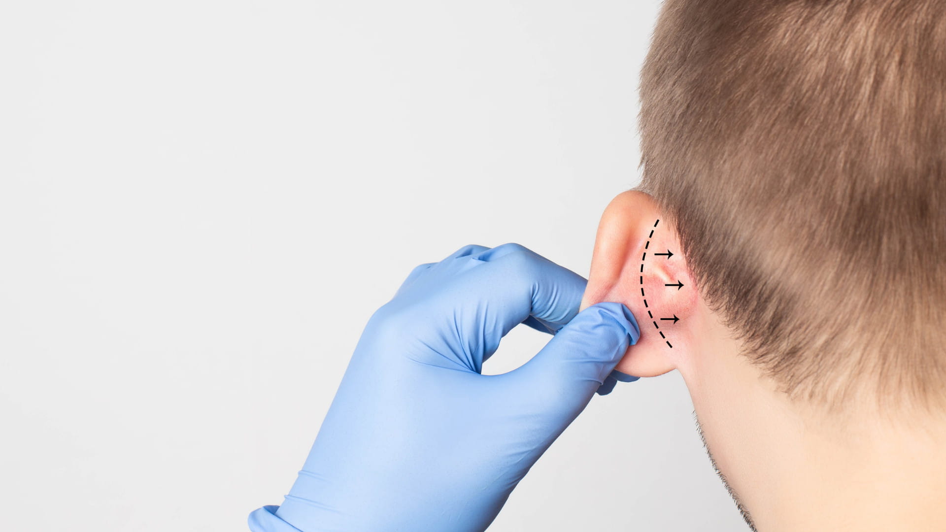
Otoplasty is a surgical procedure that reshapes, repositions, or resizes the outer ear cartilage to improve facial balance. The three primary techniques are ear pinning, ear reduction, and ear augmentation. Most patients are eligible from age five onward, with full recovery typically achieved within six weeks.
Ears rarely get attention until they do. For children teased about prominent ears or adults quietly self-conscious about asymmetry, the outer ear carries a disproportionate psychological weight. Otoplasty addresses this through precise surgical modification of cartilage structure, correcting form without altering function.
This guide breaks down every clinically relevant aspect of the procedure, from surgical subtypes and anesthesia profiling to the exact post-operative recovery protocol surgeons use to protect newly repositioned cartilage. You will also find a structured correction matrix comparing conditions, causes, and optimal treatment windows, so that you or a consulting physician can identify the appropriate intervention without ambiguity.
What Are the Three Main Types of Otoplasty?
Otoplasty is a specialized plastic surgery procedure designed to alter the size, shape, position, or structural symmetry of the outer ear cartilage, without affecting inner ear functionality or hearing mechanics. The procedure divides into three distinct surgical subtypes, each targeting a separate anatomical concern.
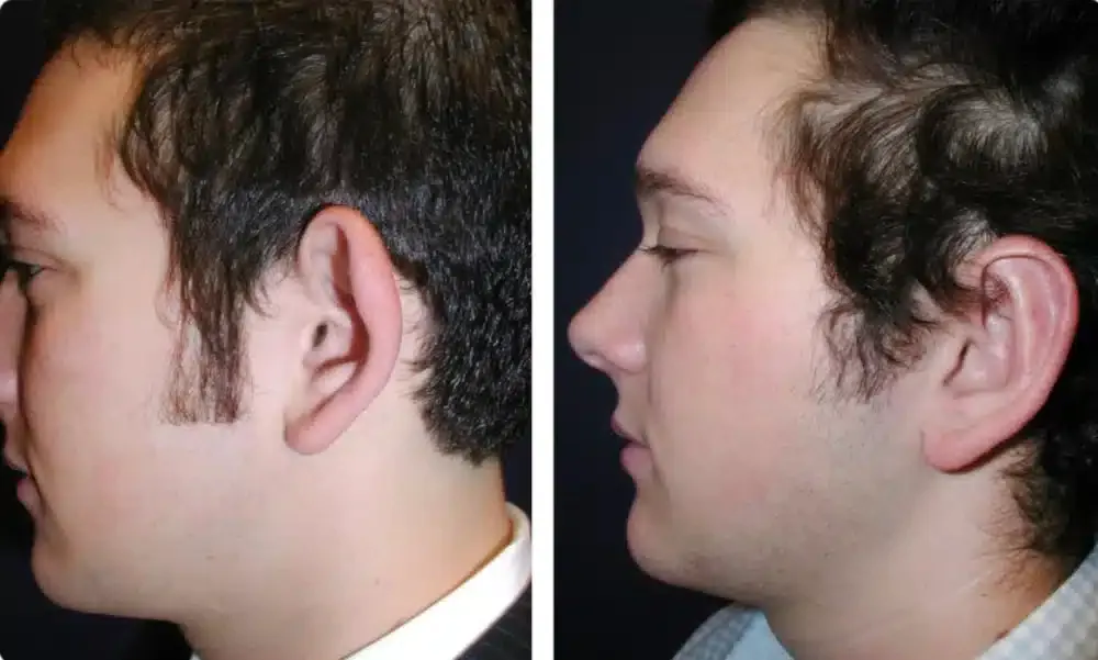
Ear Pinning (Conchal Setback)
The most frequently performed variation, conchal setback surgery corrects protruding or prominent ears by repositioning the cartilage closer to the skull. Prominence typically results from one of two structural causes: underdevelopment of the antihelical fold (the curved ridge of cartilage visible from the front) or over-projection of the conchal bowl (the deep cup-shaped cavity closest to the ear canal). Surgeons address each cause through targeted suture placement or cartilage reshaping, depending on which structural factor is dominant.
Ear Reduction (Macrotia Correction)
Macrotia refers to the clinical condition of abnormally large ears relative to facial proportions. Ear reduction surgery reduces the overall size of the outer ear framework by removing carefully calculated sections of cartilage and skin. Because the ear continues growing through adolescence, surgeons typically recommend delaying this procedure until post-puberty to ensure cartilage growth has stabilized.
Ear Augmentation for Microtia
Microtia is a congenital condition in which the outer ear is severely underdeveloped or entirely absent from birth. Surgical augmentation reconstructs the external ear structure using rib cartilage grafts harvested from the patient’s own chest wall, or alternatively, using a biocompatible implant framework. This is the most complex otoplasty subtype, often requiring multiple staged procedures over several years.
Infant Ear Molding vs. Adult Otoplasty: When Is Surgery Actually Avoidable?
Surgery is not always the first or only option. The appropriateness of a non-surgical approach depends entirely on the patient’s age at the time of diagnosis.
The Soft Cartilage Window in Newborns
During the first days and weeks after birth, elevated concentrations of maternal estrogen in a newborn’s bloodstream temporarily increase ear cartilage pliability. This biological window, which closes as maternal estrogen clears from the infant’s system (typically within the first six to eight weeks of life), allows clinicians to physically reshape malformed cartilage using non-surgical splinting molds applied directly to the ear. When treatment begins within the first seven to fourteen days of life, ear splinting achieves a high rate of structural correction without any surgical incision.
Why Standard Otoplasty Waits Until Age Five
For children who miss the newborn molding window, the next clinically appropriate time for intervention is age five. By this developmental milestone, the outer ear cartilage has reached approximately 85 to 90 percent of its adult size. Operating before this threshold risks disturbing cartilage that has not yet finished its natural growth cycle, which can lead to unpredictable long-term outcomes. Surgeons generally prefer to wait until cartilage is structurally mature enough to hold sutures reliably and maintain its repositioned form.
The Ear Correction Selection Matrix
Understanding the difference between structural anomalies helps narrow down the appropriate surgical or non-surgical intervention.
| Clinical Concern | Primary Cause | Ideal Correction Method | Optimal Treatment Window |
| Protruding / Prominent Ears | Underdevelopment of the antihelical fold or an over-projected conchal bowl | Conchal Setback (Ear Pinning Surgery) | Any time after age 5 into adulthood |
| Oversized Ears (Macrotia) | Overgrowth of the outer ear cartilage framework | Ear Reduction Surgery | Post-puberty to ensure stable growth |
| Congenital Folds (Infants) | Soft cartilage folding abnormally at birth | Non-Surgical Ear Splinting / Molding | First 7 to 14 days of life |
| Asymmetric Placement | Uneven developmental growth patterns | Unilateral or Bilateral Custom Otoplasty | Age 5 or older |
What Happens During the Otoplasty Procedure Itself?
Anesthesia: Local Sedation vs. General Anesthesia
Anesthesia selection is determined primarily by the patient’s age and capacity to remain still throughout the procedure. Adult patients typically receive local anesthesia combined with intravenous sedation, allowing them to remain conscious but fully comfortable. Pediatric patients almost universally require general anesthesia to eliminate movement risk and reduce procedural anxiety during a surgery that, depending on complexity, can last between one and three hours.
Cartilage Scoring vs. Cartilage Sparing Techniques
Once anesthesia is administered and the incision is made (typically in the natural crease behind the ear), the surgeon modifies the cartilage using one of two foundational approaches.
Cartilage scoring involves making shallow, controlled incisions or abrasions along the cartilage surface using a specialized instrument. These microscopic alterations weaken the cartilage’s structural memory at precise points, allowing the surgeon to bend and reposition it into the desired shape. This technique is often preferred when significant reshaping of the antihelical fold is required.
Cartilage sparing techniques, by contrast, involve no removal or cutting of cartilage tissue. The surgeon places permanent internal sutures (commonly Mustarde or Furnas sutures) to fold and hold the cartilage in its corrected position. This approach preserves the natural architecture of the ear and reduces tissue disruption, but its effectiveness depends on the flexibility and condition of the cartilage at hand.
Many surgeons combine both methods within a single procedure, using scoring to reshape and sutures to secure.
The Otoplasty Recovery Protocol (Step-by-Step)
Recovery from otoplasty is methodical. The primary clinical goal during the healing period is protecting the repositioned cartilage long enough for internal sutures to fully integrate and for the tissue to set permanently in its new structural position.
Step 1: Maintain the Post-Surgical Compression Wrap (Days 1 to 3)
The surgeon applies a sterile compression head dressing immediately after the procedure. This heavy wrap must remain completely dry and intact for the first several days. Its function is to prevent blood pooling (hematoma formation) beneath the delicate ear skin, which is the most common early complication of otoplasty.
Step 2: Transition to the Protective Headband (Weeks 1 to 2)
Once the primary dressing is removed at the first post-operative appointment, patients transition to wearing a soft, non-constricting headband continuously, 24 hours a day. The headband prevents the ears from accidentally folding forward, particularly during sleep, when unconscious movement poses the greatest risk to suture integrity.
Step 3: Enforce Night-Only Headband Use (Weeks 3 to 6)
Daytime headband use is discontinued, but nighttime wear continues for an additional four weeks. Sleeping flat on the back using a travel pillow or donut-shaped cervical pillow eliminates lateral pressure on the healing tissue. Any sustained compression against a pillow during this phase risks pulling the cartilage out of alignment before the sutures have fully bonded with the surrounding tissue.
Step 4: Monitor Incision Hygiene Continuously
Clean the incisions located in the crease behind the ear using a sterile cotton swab lightly moistened with saline solution. Avoid all strenuous physical activity, including contact sports, heavy lifting, and cardiovascular exercise, until the attending surgeon formally clears the patient at the six-week follow-up appointment.
Navigating Risks, Symmetry Expectations, and Post-Op Red Flags
The Symmetry Reality That Surgeons Communicate Before Every Procedure
Human ears are not naturally symmetrical. Mild variation in position, size, and contour between the left and right ear is a universal anatomical characteristic. The clinical objective of otoplasty is not identical ears but balanced facial proportions. Patients who approach surgery expecting mathematically perfect bilateral symmetry are likely to experience dissatisfaction regardless of technical outcome. Reputable surgeons establish this distinction explicitly during pre-operative consultations.
Post-Operative Complications Worth Knowing
Hematoma: A collection of blood beneath the skin surface, signaled by sudden swelling, firmness, and increasing pain in the first 24 to 72 hours. Prompt drainage by the surgical team prevents permanent deformity.
Perichondritis: A bacterial infection of the cartilage membrane (perichondrium), presenting as spreading redness, warmth, and tenderness beyond the incision site. This complication requires immediate antibiotic treatment; delayed intervention risks permanent cartilage damage.
Overcorrection: Excessive “pinned-back” positioning that creates an unnatural, flattened appearance. This typically results from overly aggressive suturing and may require revisional surgery to partially release the cartilage.
Any sign of the above warrants same-day contact with the operating surgeon.
What Every Otoplasty Candidate Should Know Before Booking a Consultation
Otoplasty produces long-term, structurally stable results when the correct technique is matched to the correct condition, performed at the clinically appropriate age, and followed by a disciplined recovery protocol. The procedure does not change hearing, does not leave visible frontal scarring, and carries a well-documented satisfaction rate among both pediatric and adult patient populations.
For anyone at the research stage, the most productive next step is a consultation with a board-certified plastic surgeon who specializes in facial procedures. Bring photographs documenting the specific concern, prepare questions about whether cartilage scoring or suturing is more appropriate for your anatomy, and ask your surgeon to define realistic symmetry expectations before the procedure date is set. While researching surgical options, understanding emotional and physical well-being—such as learning what depersonalization is, its causes, and how to stop it—can also help you maintain a balanced mindset during recovery.
Frequently Asked Questions About Otoplasty
At what age can a child have otoplasty?
Standard otoplasty is appropriate from age five onward, once the outer ear cartilage has reached approximately 85 to 90 percent of its adult size. For infants with congenital ear folding anomalies, non-surgical ear molding within the first seven to fourteen days of life is a highly effective alternative that avoids surgery entirely.
How long does otoplasty recovery take?
Most patients return to school or desk-based work within one to two weeks. Full clearance for strenuous physical activity, contact sports, and unrestricted sleep positioning is typically granted at the six-week post-operative appointment.
Does otoplasty affect hearing?
Otoplasty modifies only the outer ear (the auricle), which plays no functional role in sound transmission or hearing mechanics. The procedure does not interact with the ear canal, eardrum, or any component of the middle or inner ear, and carries no risk of hearing impairment.
What is the difference between cartilage scoring and cartilage sparing techniques?
Cartilage scoring involves making shallow abrasions in the cartilage surface to weaken its structural memory and allow reshaping. Cartilage sparing techniques use permanent internal sutures to fold and hold cartilage in position without cutting tissue. Many surgeons combine both approaches within a single procedure depending on individual anatomy.
What does otoplasty cost?
Surgical fees vary significantly by geographic region, surgeon experience, and procedural complexity. Cosmetic otoplasty is generally not covered by insurance, though procedures performed to correct congenital conditions such as microtia may qualify for partial coverage in certain health plans.
Can otoplasty be reversed?
Otoplasty is considered a permanent procedure. Overcorrection or dissatisfying results can sometimes be partially addressed through revisional surgery, but the original ear position cannot always be fully restored once sutures and scar tissue have integrated with the cartilage.
For more evidence-based articles on common health conditions, self-care, and wellness, explore our health resource library.