


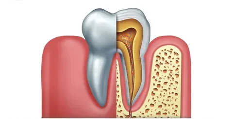
An RCT (Root Canal Treatment) is an endodontic procedure that saves a severely infected or decayed tooth. The rcts dental removes the damaged pulp, disinfects the root canals, and seals them with gutta-percha to block future bacterial invasion. Most patients report little to no pain during the procedure itself.
Root canals have an undeserved reputation. Ask most people how they feel about getting one, and you’ll hear words like “terrifying” or “painful.” The reality? Modern root canal treatment is closer to a routine filling than the horror story passed around at dinner tables. The discomfort most patients associate with an RCT is actually the infection that made the treatment necessary, not the procedure itself.
So what actually happens during a root canal? This guide walks through every clinical stage in plain language, covering the instruments used, the reasoning behind each step, and what makes the difference between a root canal that lasts decades and one that fails within a year.
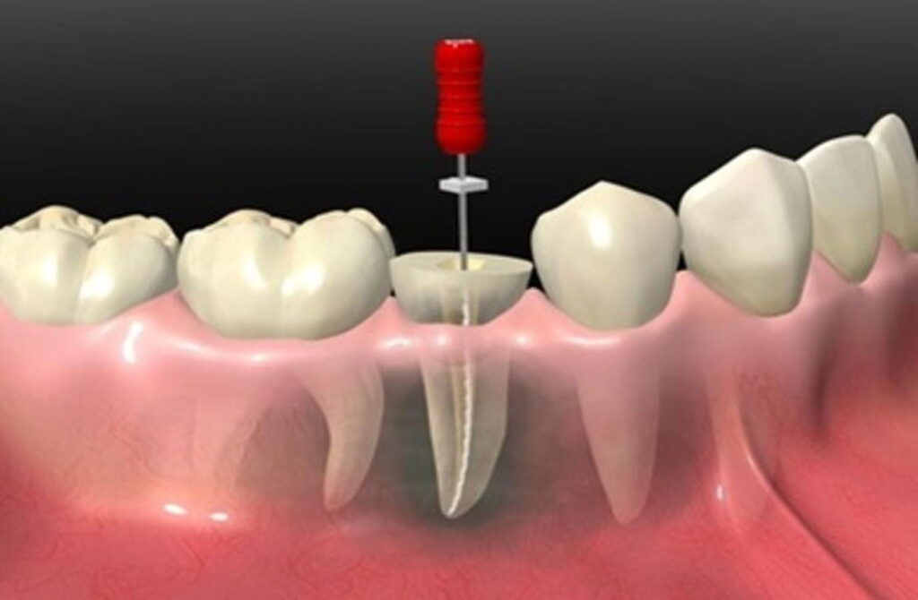
What Is Root Canal Treatment (RCT) in Dentistry?
Root canal treatment is a specialized endodontic procedure designed to save a tooth that would otherwise require extraction. Deep decay, a dental abscess, a cracked tooth, or repeated dental procedures on the same tooth can all destroy the soft tissue living inside the tooth root. That soft tissue, called the dental pulp, contains nerves, blood vessels, and connective tissue. Once it becomes severely infected or dies, it must be removed.
The procedure involves cleaning out that infected pulp, disinfecting the hollow root canals left behind, and sealing the entire system to prevent bacteria from re-entering. After treatment, the tooth can continue functioning normally for the rest of the patient’s life, provided it receives a proper protective crown.
What Are the Six Clinical Stages of Root Canal Treatment?
Understanding the physical sequence of an RCT removes much of the fear surrounding the procedure. Each stage has a specific purpose, and skipping or rushing any one of them compromises the final outcome.
Stage 1: Access Cavity Preparation
The dentist drills through the biting surface of the tooth using high-speed diamond burs to create a direct opening into the pulp chamber. This access point must be precisely positioned. A poorly placed access cavity forces endodontic instruments to bend at awkward angles, which increases the risk of file breakage inside the canal.
Stage 2: Canal Shaping
Rotary nickel-titanium (NiTi) files are inserted into each root canal to remove infected dentin from the canal walls and gradually widen the space. NiTi files are preferred over stainless steel because the alloy flexes through curved root anatomy without snapping. Canal shaping creates a smooth, uniform taper that allows irrigating fluids to penetrate evenly and makes the final sealing step far more predictable.
Stage 3: Chemical Irrigation and Disinfection
Mechanical filing alone cannot reach every corner of a root canal system. Many teeth contain microscopic lateral canals that branch off the main root. These spaces harbor bacteria and organic debris that physical instruments simply cannot contact.
Sodium hypochlorite (NaOCl), the same compound found in household bleach but at a clinical concentration, is flushed repeatedly through the canals to dissolve organic matter and kill bacteria. A chelating agent called EDTA follows to remove the smear layer left behind by the rotary files. Ultrasonic activation is often used to agitate these solutions and force them deeper into lateral branches.
Stage 4: Obturation with Gutta-Percha
Once the canals are confirmed sterile and completely dry, they are sealed with gutta-percha. Gutta-percha is a biocompatible, rubberized material derived from the sap of the Palaquium tree. Custom-fit gutta-percha cones are coated in a resin sealer cement and condensed tightly into the root system using heat.
This hermetic seal is the procedure’s primary defense against re-infection. Any gap or void left in the obturation gives oral bacteria a pathway back into the root and, from there, into the surrounding jawbone.
Stage 5: Core Buildup
After the roots are sealed, the dentist rebuilds the structural core of the tooth crown using composite resin or glass ionomer cement. The access cavity created at the start of the procedure, combined with the decay that caused the infection, often leaves very little natural tooth structure remaining. The core buildup restores enough volume to support the final restoration.
Stage 6: Crown Placement
A full-coverage porcelain or zirconia crown is placed over the treated tooth, typically within a few weeks of completing the root canal. This stage is not optional, and the biology behind it explains why.
Why Does a Root Canal Tooth Need a Crown?
A common point of patient confusion is why a crown is required after an RCT when the tooth already appears intact. The answer lies in what the dental pulp actually does for a living tooth.
The pulp supplies continuous moisture and nutrients to the surrounding dentin matrix. When the pulp is removed, the dentin gradually dries out over the following months. Dehydrated dentin becomes brittle, far more susceptible to fracture under normal chewing forces than a vital, living tooth. A vertical root fracture, the most common failure mode in uncrowned post-RCT teeth, cannot be repaired. The tooth must be extracted immediately.
A protective crown distributes chewing forces evenly across the tooth and effectively eliminates this fracture risk. Patients who skip this final step often lose the tooth they went through the procedure to save.
What Happens During Each Phase of the Procedure in the Chair?
The clinical appointment follows a clear sequence that keeps the procedure sterile and precise from start to finish.
Isolation. Before any instrument enters the tooth, the clinician applies a dental dam: a thin rubber sheet that isolates the tooth from the rest of the mouth. This step is non-negotiable. Saliva carries millions of bacteria, and a single contamination event during canal preparation can reintroduce infection into a canal that was just cleaned.
Working length determination. A digital apex locator measures the exact millimeter length of each root canal down to its terminal tip, called the apex. Confirmation X-rays verify the measurement. Cleaning instruments that travel past the apex can damage the periapical tissue at the root tip, triggering inflammation, pain, and delayed healing.
Deep chemical debridement. Multiple rounds of irrigating solution are cycled through the canals between each file size. The volume and frequency of irrigation matter considerably. Research in endodontics consistently identifies inadequate irrigation as one of the leading causes of root canal failure.
Packing and condensation. The gutta-percha cones are fitted, coated in sealer, and heat-condensed to fill every millimeter of the prepared canal space. A post-obturation X-ray confirms that the seal reaches the apex without overfilling beyond it.
How Long Does Root Canal Treatment Take?
Most root canal procedures are completed in one or two appointments, each lasting between 60 and 90 minutes. Molars have three or four root canals compared to the single canal in a front tooth, so posterior teeth generally require more time. Teeth with prior treatment, curved roots, or calcified canals take longer to negotiate safely.
Root Canal Treatment FAQs
Is root canal treatment painful?
Most patients report that the procedure itself is no more uncomfortable than a routine filling. The tooth and surrounding tissue are fully anesthetized before any instruments are used. Soreness for two to three days after the appointment is normal and typically responds well to over-the-counter anti-inflammatories.
How long does a root canal treated tooth last?
A tooth with a successful RCT, a well-adapted crown, and good oral hygiene can last a lifetime. Studies published in the Journal of Endodontics report survival rates exceeding 90% at 10 years when the tooth is properly restored with a crown following treatment.
What are the signs that a tooth might need root canal treatment?
Common indicators include prolonged sensitivity to heat or cold, spontaneous toothache, tenderness when biting, darkening of the tooth, and swelling or a persistent pimple on the gum near the tooth root. An X-ray can often reveal an abscess or bone loss at the root tip before symptoms become severe.
What is the difference between an RCT and a tooth extraction?
Root canal treatment saves the natural tooth. Extraction removes it entirely, which then requires a dental implant, bridge, or partial denture to restore function and prevent neighboring teeth from shifting. Keeping the natural tooth is almost always the preferred clinical outcome when it remains viable.
Can a tooth that has already had a root canal become infected again?
Yes. Re-infection can occur if the original obturation was incomplete, if the crown seal breaks down over time, or if a new cavity develops. This situation requires a retreatment procedure, where the original gutta-percha is removed, the canals are cleaned again, and the tooth is resealed.
The Bottom Line on Root Canal Treatment
Root canal treatment is one of the most misunderstood procedures in dentistry. The six-stage process, from access cavity to protective crown, is designed around a single goal: eliminating infection completely and sealing the tooth so permanently that bacteria cannot return.
The procedure’s reputation for pain belongs to a different era of dentistry. Contemporary anesthetic techniques, rotary NiTi instrumentation, and digital apex locators have turned RCT into a predictable, comfortable, and highly successful treatment. If your dentist or endodontist has recommended one, the tooth is almost certainly worth saving. Acting sooner rather than later preserves more natural tooth structure, simplifies treatment, and typically results in a better long-term outcome—just as taking timely action after major life changes can help you explore restoration options like a breastfeeding lift and post-nursing mastopexy guide for personal health and confidence.
Continue exploring expert-written articles on everyday health topics by visiting our health knowledge center.